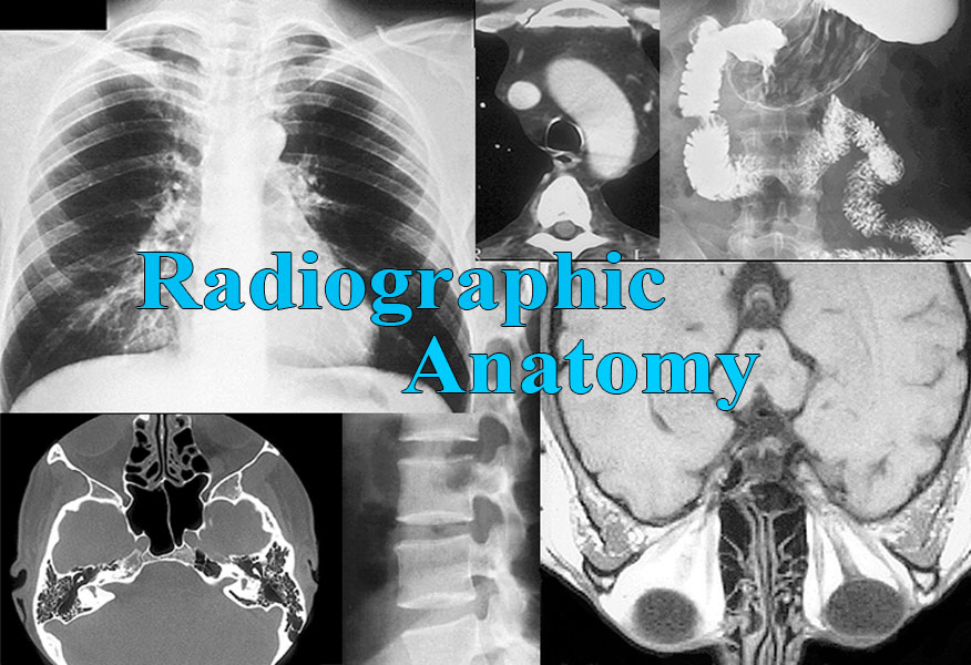


The study of radiographs is highly dependent on one's knowledge of gross anatomy. It is fair to say that a conventional x-ray is essentially a two-dimensional, black-and-white image of a patient's gross anatomy.
Your approach to studying the images of Radiographic Anatomy depends on the context of your use of the images. If you are enrolled in or have previously completed an anatomy course that describes the gross anatomy of the human body, that course content should serve you well in appreciating the anatomy seen in the radiographs. In this case the Correlation image(s) will re-enforce the gross anatomy being or having been learnt previously. In contrast, if you do not have an appreciation for the gross anatomy of the body regions in the radiographs, you may want to study the Correlation image(s) that accompany a radiograph before looking at the radiograph itself. This approach will provide a better appreciation for the anatomy that will be described in the radiograph.
There is also a logic to the organization of the images in the menus; the images were not randomly positioned within the menus. For example, the Upper Limb and Lower Limb radiographs are arranged in their menus from the proximal end of the limb to its distal end; the same sequence in which a study of the gross anatomy of the limbs would typically occur. The Abdomen menu is organized by organ systems, the topic of angiography, and so on. If your intent is to study all of the radiographs in a body region, we suggest that you begin your study with the first image in the series and work your way sequentially to the last image.