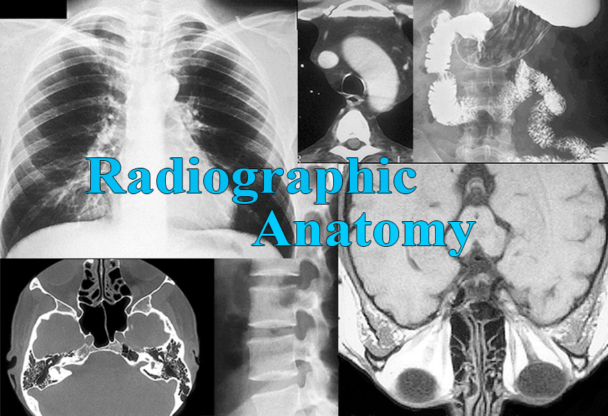


The Radiographic Anatomy portion of NetAnatomy is designed to teach and reinforce an appreciation for human anatomy using a visual medium, radiology, that is common to the health care professions. The various radiological modalities, e.g. plain film, MRI, etc. provide visually unique representations of human anatomy that supplement other types of anatomical observation, e.g. cadaveric specimens. Radiological images are generally derived from living subjects and, while one must appreciate the inherent limitations of the radiographs themselves, they are free of postmortem artifacts. One need not desire to be a radiologist in order to benefit from a study of radiological images; they provide valuable and interestingly depicted information on the positions, sizes, shapes, and relationships of organs and structures.
Radiology is the branch of medicine concerned with the diagnosis and treatment of disease utilizing techniques that employ both ionizing (e.g. x-rays) and nonionizing (e.g. ultrasound) radiation. For students of the health care professions Radiographic Anatomy provides an appropriate introduction to radiology without losing site of the primary mission of NetAnatomy, i.e. to facilitate study of the structure and function of the human body. Since anatomical study is the primary goal of NetAnatomy, emphasis is placed on the normal anatomy represented in the radiographic images. However, pathology can be defined in part by a deviation from the normal; thus the normal anatomy learnt in Radiographic Anatomy will serve as a reference point for those who pursue professions that address the diagnosis and treatment of disease.